Home » Without Label » Anatomical Name Of Lower Back Muscles - Anatomical Name Of Lower Back Muscles - Amazon Com Labeled ... / Balance the weight of your head on top of your spine
Anatomical Name Of Lower Back Muscles - Anatomical Name Of Lower Back Muscles - Amazon Com Labeled ... / Balance the weight of your head on top of your spine
Anatomical Name Of Lower Back Muscles - Anatomical Name Of Lower Back Muscles - Amazon Com Labeled ... / Balance the weight of your head on top of your spine. And reach, pull and extend your arms and torso. This blog post article is an overview of the muscles of the lumbar spine of the trunk. The back anatomy includes some of the most massive and functionally important muscles in the human body. Bones of the pelvis and lower back. This curve, called lordosis, helps to:
Sherwin is a medical research scientist and author of the low back pain program and ebook. In the meanwhile, your hip flexors, quadriceps and lumbar muscles remain tight to keep you in an upright position. The vertebral column of the lower back includes the five lumbar vertebrae, the sacrum, and the coccyx. Intermediate back muscles and c. Still, many individuals pay far too little attention to them.
Pin on human anatomy drawing from i.pinimg.com The l5 vertebra is connected to the top of. The muscle then courses up to your shoulder and attaches to your upper arm bone. The quick answer to this question is the muscles of the lower back are the multifidus, longissimus, spinalis, and quadratus lumborum. In this image, you will find an occipital bone, sternocleidomastoid, trapezius, deltoid in muscles of the lower back diagram. The psoas muscle is a low back muscle located deep in the body, very close to the spine and inside the hip and thigh bones. This curve, called lordosis, helps to: The gastrocnemius is connected to the heel by the achilles tendon. Its name means belly of the leg,and its common name is the calf muscle.
As you can see, there are also have a spine of scapula deltoid, triceps brachii, latissimus dorsi.
With over 20 years of research experience from the toronto general hospital and the hospital for sick children, he provides sensible, effective, advice and solutions for lower back pain. They originate from the thoracolumbar fascia, the spinous process of thoracic six through 12, the iliac crest, and your lower three ribs. These muscles include the large paired muscles in the lower back, called erector spinae, which help hold up the spine, and gluteal muscles. There are three parts to the trapezius. Sherwin is a medical research scientist and author of the low back pain program and ebook. They help to bend the back to one side or the other. The lumbar spine is the lower back that begins below the last thoracic vertebra (t12) and ends at the top of the sacral spine, or sacrum (s1). This curve, called lordosis, helps to: The quadratus lumborum muscles (orange, in the image above) are found in the lower back (also called the lumbar area). The l5 vertebra is connected to the top of. Muscles of lower back diagram. The muscles of the lower back, including the erector spinae and quadratus lumborum muscles, contract to extend and laterally bend the vertebral column. L1, l2, l3, l4, and l5.
In this image, you will find an occipital bone, sternocleidomastoid, trapezius, deltoid in muscles of the lower back diagram. The psoas muscle is a low back muscle located deep in the body, very close to the spine and inside the hip and thigh bones. The lordotic curve your lower back (lumbar spine) is the anatomic region between your lowest rib and the upper part of the buttock. The trapezius or trapezoid muscles are two paired muscles that extend from the base of the thoracic vertebrae in the spine to the occipital bone and run out to the spine of the scapula. The quadratus lumborum muscles (orange, in the image above) are found in the lower back (also called the lumbar area).
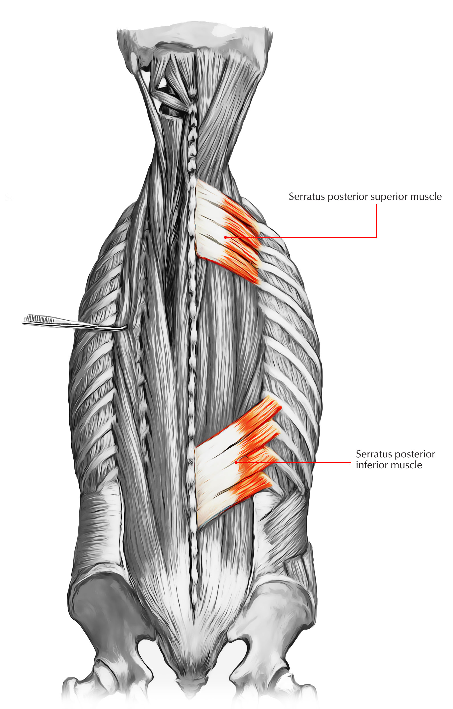
FreeFitnessGuru - Rear Leg Anatomy from www.freefitnessguru.com The l5 vertebra is connected to the top of. The gastrocnemius runs down the back of the lower leg, from the end of the femur to the heel bone, or calcaneus. The superficial back muscles include the suboccipital muscles, trapezius, latissimus dorsi, levator scapulae, rhomboids and serratus posterior muscles. Anatomical name of lower back muscles : The quadratus lumborum muscles (orange, in the image above) are found in the lower back (also called the lumbar area). The posterior superficial muscles are the three gluteal muscles (gluteus maximus, gluteus medius, gluteus minimus), and the tensor fascia latae. In turn, the posterior deep muscles are the piriformis, obturator internus, obturator externus, superior gemellus, inferior gemellus, and quadratus femoris. In this image, you will find an occipital bone, sternocleidomastoid, trapezius, deltoid in muscles of the lower back diagram.
With over 20 years of research experience from the toronto general hospital and the hospital for sick children, he provides sensible, effective, advice and solutions for lower back pain.
L1, l2, l3, l4, and l5. 1 your spine in this region has a natural inward curve. The back muscles can be three types. The superficial back muscles include the suboccipital muscles, trapezius, latissimus dorsi, levator scapulae, rhomboids and serratus posterior muscles. The muscles of the back that work together to support the spine, help keep the body upright and allow twist and bend in many directions. The quadratus lumborum muscles (orange, in the image above) are found in the lower back (also called the lumbar area). The vertebral column of the lower back includes the five lumbar vertebrae, the sacrum, and the coccyx. See back muscle anatomy stock video clips. These structures work together to support the body, enable a range of movements, and send messages from the brain to. When it contracts, it makes the foot bend downward, and it also helps to bend the knee. With over 20 years of research experience from the toronto general hospital and the hospital for sick children, he provides sensible, effective, advice and solutions for lower back pain. Lumbar spine lower back and superficial muscles the muscles of the lower back help stabilize, rotate, flex, and extend the spinal column, which is a bony tower of 24 vertebrae that gives the body. The trapezius or trapezoid muscles are two paired muscles that extend from the base of the thoracic vertebrae in the spine to the occipital bone and run out to the spine of the scapula.
The quadratus lumborum muscles (orange, in the image above) are found in the lower back (also called the lumbar area). Bones of the pelvis and lower back. The anatomy of the lumbar spine is quite complex. See back muscle anatomy stock video clips. These muscles provide posture and stability to the body by holding the vertebral column erect and adjusting the position of the body to maintain balance.
Back Muscles - 28 Major 【Muscles of the Back】 - Earth's Lab from www.earthslab.com The psoas muscle is a low back muscle located deep in the body, very close to the spine and inside the hip and thigh bones. Muscles of the shoulder 12 photos of the muscles of the shoulder muscles of the neck shoulder and back, muscles of the outer shoulder, muscles of the thorax shoulder and abdominal wall, muscles over shoulder blades, posterior muscles of the neck shoulder and back, human muscles, muscles of the neck shoulder and back, muscles of … Let us introduce you to each of these muscles presented in our diagram. Anatomical name of lower back muscles : The muscles of the lower back, including the erector spinae and quadratus lumborum muscles, contract to extend and laterally bend the vertebral column. When it contracts, it makes the foot bend downward, and it also helps to bend the knee. (2017, elsevier) should be consulted. Related posts of muscle names of lower back cadaver muscle anatomy.
Muscles of the lumbar spine.
The quadratus lumborum muscles (orange, in the image above) are found in the lower back (also called the lumbar area). Human musculature bodybuilding infographic muscular system vector human anatomy back muscle anatomy bicep male muscular anatomy human body anatomy female female anatomy muscle hamstrings muscle. L1, l2, l3, l4, and l5. The lumbar spine makes up the the lower end of the spinal column. Here we will attempt to provide a brief overview of lumbar spinal anatomy. The back consists of the spine, spinal cord, muscles, ligaments, and nerves. The muscles of the back that work together to support the spine, help keep the body upright and allow twist and bend in many directions. The bones of the pelvis and lower back work together to support the body's weight, anchor the abdominal and hip muscles, and protect the delicate vital organs of the vertebral and abdominopelvic cavities. These muscles provide posture and stability to the body by holding the vertebral column erect and adjusting the position of the body to maintain balance. In the meanwhile, your hip flexors, quadriceps and lumbar muscles remain tight to keep you in an upright position. It is composed of trapezius, latissimus dorsi, rhomboid major, rhomboid minor and levator scapulae. Still, many individuals pay far too little attention to them. And reach, pull and extend your arms and torso.